의료장비 |
|
연구장비 |
CK피부분석 및 상태측정장비, 생체나이측정장비 |
피부상태 검사용 광학측정장비 [SkinDex 300][VivaScope 1500]
| VivaScope 1500 | ||
|
The VivaScope?/FONT> 1500. Loaded with features and effortless to use.
Non-invasive cellular imaging.
Results you've never seen before.
VivaScopysm provides
a 'window' into tissue for cellular imaging of skin and other living
tissues. The technology is entirely non-invasive and images naturally
occuring index of refraction differences in tissue. The non-invasive
nature of a VivaScope?nbsp;'optical biopsy' allows repeated imaging
of a single tissue site. This enables monitoring natural biological
processes in-vivo, over time. Only Lucid's VivaScopes?nbsp;provide
video rate in-vivo confocal images that allow visualization
of real-time processes such as blood flow in capillary loops and monitoring
of leukocyte trafficking in skin.
Imaging living tissue
provides the key to understanding.
Using the VivaScope?/FONT>
1500 cellular imager you can readily observe cellular and nuclear features
in skin or other exposed living tissue, one cell layer at a time.
Video rate images of thin virtual sections of skin are easily obtained,
in real-time, without the need for an invasive biopsy. The understanding
gained through Vivascopysm can save time and money in clinical
testing, get products to market faster and provide increased confidence
for a successful new product launch.
|
 The
VivaScope?/FONT>
1500 is a second generation in-vivo laser confocal microscope
capable of imaging living tissue at the cellular level. The VivaScope?/FONT>
1500 has been designed specifically to meet the stringent demands of
researchers investigating living processes at the cellular level.
It has the features needed for basic scientific and clinical research
of skin and other tissues. The 1500 provides higher resolution,
improved ergonomics and more features, all at a lower cost than was
possible with the previous generation of instruments.
The
VivaScope?/FONT>
1500 is a second generation in-vivo laser confocal microscope
capable of imaging living tissue at the cellular level. The VivaScope?/FONT>
1500 has been designed specifically to meet the stringent demands of
researchers investigating living processes at the cellular level.
It has the features needed for basic scientific and clinical research
of skin and other tissues. The 1500 provides higher resolution,
improved ergonomics and more features, all at a lower cost than was
possible with the previous generation of instruments.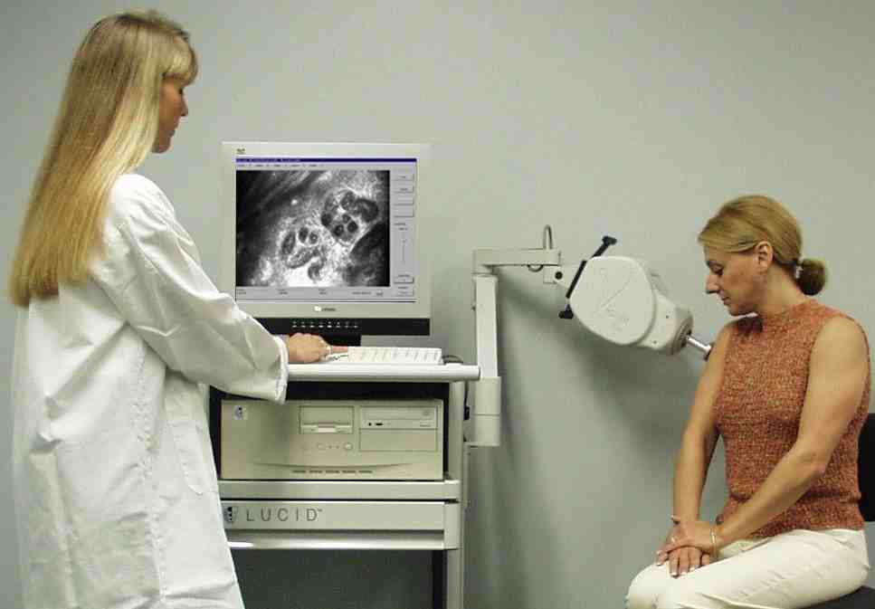 The VivaScope?/FONT>
1500 has a gimbal mounted imaging head and multi-axis arm to allow precise
placement of the confocal objective while simultaneously providing a
high degree of patient comfort. Set-up of the imaging session
is aided by the control keypad located on the imaging head. The
1500 uses Lucid's proprietary StableViewtm(tm) confocal microscope
objectives and patented thick window tissue rings. These provid
an unprecedented degree of tissue stabilization, allowing generation
of precision and repeatable image stacks and maps. The 19' flat
panel display and megapixel confocal optics define the state-of-the-art
for real-time in-vivo image resolution. No other system
provides this combination of features in a low cost, easy to use
in-vivo microscope.
The VivaScope?/FONT>
1500 has a gimbal mounted imaging head and multi-axis arm to allow precise
placement of the confocal objective while simultaneously providing a
high degree of patient comfort. Set-up of the imaging session
is aided by the control keypad located on the imaging head. The
1500 uses Lucid's proprietary StableViewtm(tm) confocal microscope
objectives and patented thick window tissue rings. These provid
an unprecedented degree of tissue stabilization, allowing generation
of precision and repeatable image stacks and maps. The 19' flat
panel display and megapixel confocal optics define the state-of-the-art
for real-time in-vivo image resolution. No other system
provides this combination of features in a low cost, easy to use
in-vivo microscope.